Case A
AAPSP September 06 Canine skin-Demidecosis.jpg (98 KB)
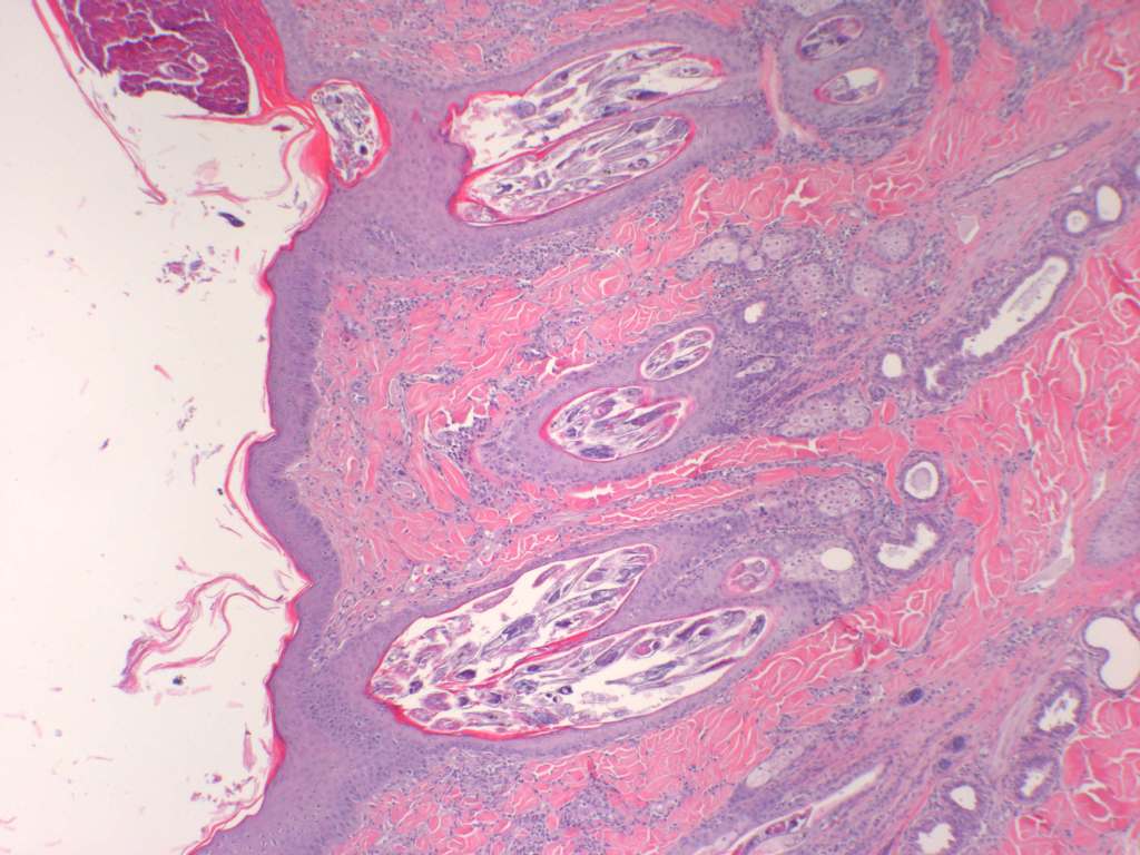
History
Mature dog with crusty skin lesions along the dorsum.
Notes on histopathological description
A description of ‘intrafollicular organisms consistent with mites’ is probably satisfactory for this common disease. However, a brief description of the distinguishing anatomical features is recommended as a general practice when parasites are present in tissue sections.
Morphological Diagnosis
Folliculitis and furunculosis, pyogranulomatous, chronic, diffuse, severe, with intralesional arthropod parasites and coccoid bacteria
Aetiological Diagnosis
Generalised pustular demodicosis. In this case the severity of furunculosis warrants designation as the pustular form of generalised demodicosis.
Comments
This is a common disease with few differentials. However, characterisation of the coccoid bacteria using a gram stain is recommended. Recovery of parasites for specialist microscopy is required for definitive identification.
Case B

AAPSP September 06 Bovine brain-Pompes disease.jpg (89 KB)
History
Neurological signs were apparent in a nine-month-old Angus bull.
Notes on histopathological description
This is a section of the medulla oblongata approximately at the level of the obex. The key features are vacuolar distension of neuronal perikarya, axonal spheroid formation and vacuolation of vascular myocytes. Gliosis is equivocal.
Morphological diagnosis
Encephalopathy, vacuolar, neuronal, chronic, diffuse, severe
Aetiological diagnosis
Lysosomal storage disease, probably generalised glycogenosis type II.
Comments
The preferred diagnosis is lysosomal storage disease, most probably generalised glycogenosis type II. The differential diagnosis should include other known genetically transferred lysosomal storage diseases (e.g. mannosidosis) and toxic diseases (e.g. swainsonine-induced mannosidosis). A novel lysosomal storage disease can not be completely excluded. Special stains for glycogen are recommended to distinguish glycogenosis from mannosidosis. Genetic testing may be needed for confirmation of glycogenosis.