Case A
AAPSP March 06 Porcine heart-Mulberry Heart disease.jpg (138 KB)
History
Weaner pig in good body condition, with a history of sudden death.
Example histopathological description
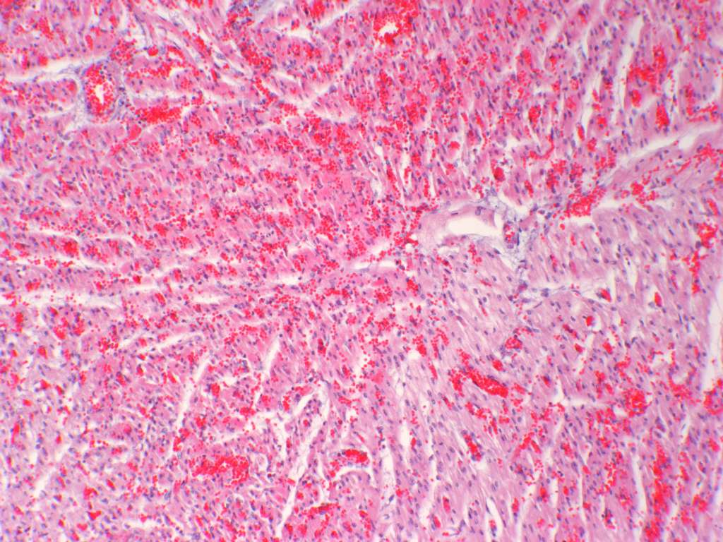
This is a transmural section of heart including epicardium and endocardium. There is multifocal and diffuse, in places severe, haemorrhage throughout the myocardium and beneath endocardium. Congestion of vessels is also apparent and oedema is manifested by marked separation of myocardial fibres, presumably by proteinaceous fluid which is seen in some areas, sometimes with fibrin strands present. Especially in association with severe haemorrhage, there is vacuolation and loss of striation of myocardial fibres, and increased eosinophilia of sarcoplasm – indicative of myonecrosis. Many polymorphonuclear neutrophils and fewer mononuclear cells are scattered throughout these areas and fibrin accumulations in some vessels are suggestive of thrombosis.
Morphological diagnosis
Cardiomyopathy, haemorrhagic and necrotising, acute, multifocal and diffuse, severe.
Aetiological diagnosis
Probable Vitamin E/ Selenium deficiency complex (mulberry heart disease).
Comments
History of illness in this weaner and other pigs in the herd, and other pathological findings should eliminate most other possible diagnoses. To confirm Vitamin E/Selenium deficiency complex as the cause, appropriate tests could include analysis of rations for content of selenium, vitamin E and polyunsaturated fatty acids, examination of liver for concentration of selenium, and analysis of blood or plasma from in-contact live pigs for glutathione peroxidase activity.
Case B

AAPSP March 06 Porcine heart-Mulberry Heart disease.jpg (138 KB)
History
Aged Merino ewe in poor body condition.
Example histopathological description
This is a section of lymph node and associated perinodal adipose tissue and vessels. The architecture of the node, especially in the cortex, is markedly altered by pathological change. Extensive, ill-defined areas of necrosis are apparent, while in other areas, especially in, but also extending out from lymph node sinuses, there are prominent accumulations of histiocytes and occasional multinucleated giant cells. Moderate to marked accumulations of lymphocytes are associated with these granulomas, and plasma cells and small foci of polymorphonuclear neutrophils are also present in some areas. Changes prominent in perinodal locations include lympho-granulomatous perivasculitis and vasculitis with thrombosis. Examination of Ziehl Neelsen-stained sections revealed only occasional small groups of acid-fast bacilli.
Morphological diagnosis
Lymphadenitis, granulomatous necrotizing, chronic-active, diffuse, severe, with lymphangitis.
Aetiological diagnosis
Acid fast bacterial infection; probable ovine Johne’s Disease caused by infection with Mycobacterium avium subsp. paratuberculosis.
Comments
Knowledge of flock history and other pathological findings in this ewe and other, in-contact, animals will rule out most other possible diseases as a cause of the observed lesions. If important, (e.g. for epidemiological or disease control reasons), a more precise diagnosis may be confirmed by conventional culture, radiometric culture, PCR or ELISA.