Case A
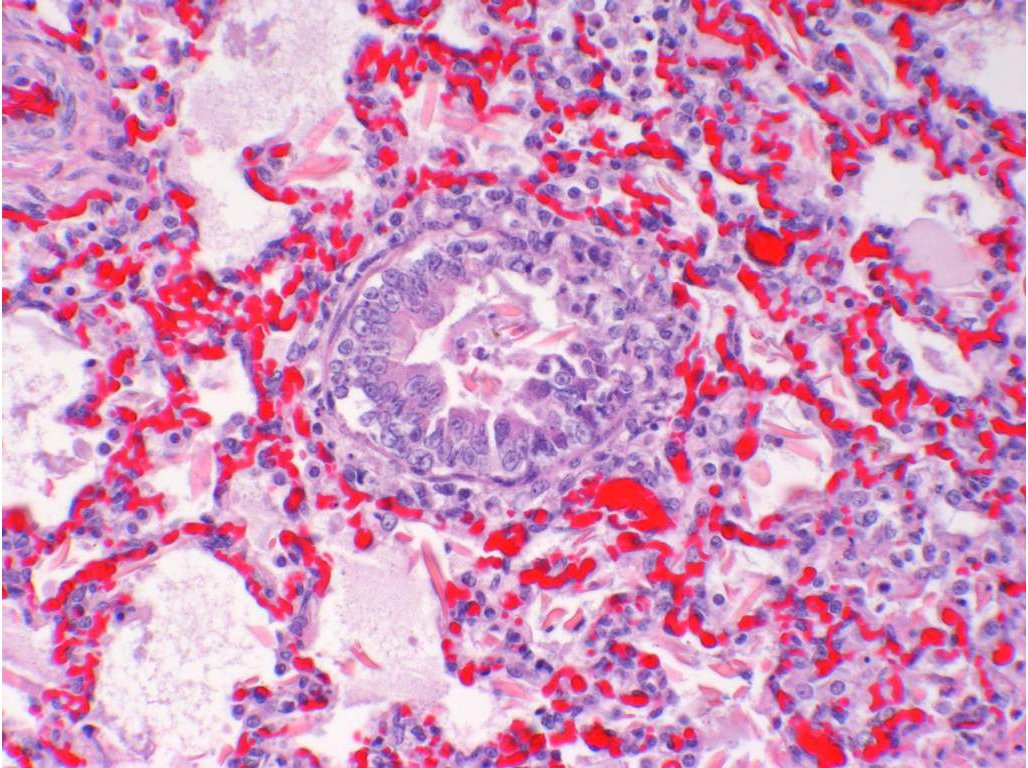
Equine foetal lung with necrotising bronchopneumonia (EHV1 infection) (196 KB)
History
Lung tissue from an aborted foal
Example Histopathological Description
This section of lung shows a fairly uniform sub-gross appearance with diffuse congestion and an increase in cellularity accompanied by thickening of the pleura and interlobular septae. The pleura and interlobular septae are oedematous and contain patchy haemorrhage and a low grade inflammatory infiltrate that consists primarily of mononuclear cells. Alveolar septae are thickened due to marked congestion and the presence of inflammatory cells. There is widespread exudation of fibrin and cells, primarily macrophages with lesser numbers of neutrophils and occasionally erythrocytes, into alveoli. Epithelial squames and necrotic debri are also common within alveoli, the former indicating foetal distress. In patchy areas there is necrosis of the alveolar septae and in others there is hyperplasia of alveolar type II cells, indicating attempts at repair. There are also multifocal areas of bronchiolar epithelial hyperplasia and necrosis, and the presence of cellular exudate within bronchiolar lumena. Numerous intranuclear, eosinophilic, inclusion bodies are present within epithelial cells of the alveolar wall and bronchioles.
Morphological diagnosis
Pneumonia, necrotising and suppurative, subacute, diffuse, severe with eosinophilic intranuclear inclusion bodies
Aetiological diagnosis
Equine herpesvirus foetal pneumonia
Aetiology
Equine Herpesvirus Type 1 (EHV1)
Comment
The changes present are characteristic of EHV-1 infection in equine foetuses. This infection should be confirmed by EHV-1 PCR on fresh lung, thymus or spleen to exclude the rare case of equine abortion due to EHV4, although abortion due to EHV4 has not been reported in Australia. EHV1 abortion is a notifiable disease in most States of Australia. EHV-1 infection, causing abortion, is highly contagious and strict control procedures, based on the guidelines provided by Equine Veterinarians Australia (formerly Australian Equine Veterinary Association), are essential in minimising spread to pregnant mares within and between stud farms. These guidelines are available on request. Abortion occurs in mares either as a result of recrudescence of a carrier state in a pregnant mare, or exposure of naive pregnant mares to infected secretions from another horse. Australian serological surveys show up to 40% of Thoroughbred horses have been exposed to the virus and are therefore considered carriers.
Case B
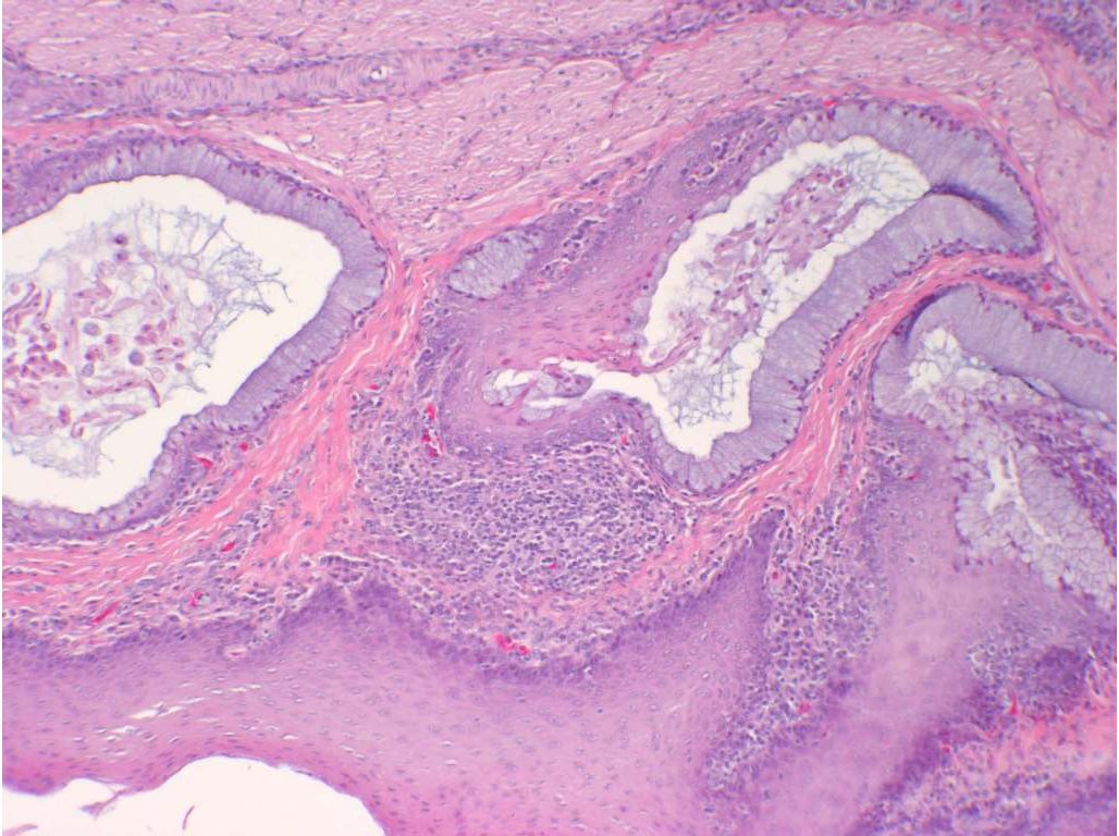
Chicken oesophagus with squamous metaplasia and inflammation of the submucosal glands-Vitamin A deficiency (162 KB)
History
Game birds showing poor growth and oculo-nasal discharge.
Example histopathological description
Three cross-sections of oesophagus are examined. There is extensive squamous metaplasia of the submucosal mucous secreting glands with frequent cystic accumulation of necrotic heterophils, mucus and/or sloughed squamous epithelial cells within affected glands. The squamous metaplasia is segmental in some glands. Some contain poorly visualised irregular purple structures that could be clumps of bacteria amongst the coagulated necrotic heterophils. In the submucosa there is variable inflammation from a light mononuclear cell or heterophilic infiltrate to an intense lymphoplasmacytic and heterophilic response. There is hyperplasia of the squamous epithelium lining the oesophagus. No viral inclusions, parasites, protozoa or fungal elements can be seen.
Morphological diagnosis
Oesophagitis, lymphoplasmacytic and heterophilic, chronic-active, diffuse, severe, with squamous metaplasia of oesophageal glands.
Aetiological diagnosis
Vitamin A deficiency
Comment
This metaplastic change is relatively characteristic for hypovitaminosis A. The mild inflammation is most likely due to secondary proliferation and invasion by bacteria. Further proof of hypovitaminosis A could be obtained from the history, nutritional analysis of the diet and determinations of levels of vitamin A in plasma and liver. For the biochemical estimations, the plasma and liver should be snap-frozen and protected from ultraviolet light.